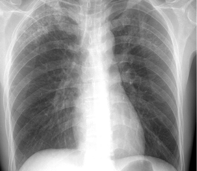
Tuberculosis X Ray / X Ray Of The Lungs Fibrous Cavernous Tuberculosis Negative Stock Photo Image Of Cavity Breast 137206508 / The lungs are the most common site of primary infection by tuberculosis and are a major source of spread of the disease and of individual morbidity and mortality.
Tuberculosis X Ray / X Ray Of The Lungs Fibrous Cavernous Tuberculosis Negative Stock Photo Image Of Cavity Breast 137206508 / The lungs are the most common site of primary infection by tuberculosis and are a major source of spread of the disease and of individual morbidity and mortality.. We also attempt to categorize the findings into those definitive for active tb, indeterminate for disease activity, and those indicating healed tb. Metrohealth and case western reserve university, affiliated since 1914, partners in advancing patient care through research and teaching. The article reviews the imaging findings in ctb on various modalities. Diagram of a treatment algorithm for active tuberculosis. A positive tb skin test or tb blood test only tells that a person has been infected with tb bacteria.
Less than 50% of adults with primary tuberculosis. It does not tell whether the person has latent tb infection (ltbi) or has progressed to tb disease. The typical image is a homogeneous or heterogeneous white spot at the base of the affected lung or the middle third. The samples are tested for tb bacteria. The main tb bacterium is mycobacterium tuberculosis (m.
Artificial Intelligence Could Help Diagnose Tuberculosis In Remote Regions Study Finds Scope from scopeblog.stanford.edu A general discussion of tuberculosis is found in the parent article: Diagram of a treatment algorithm for active tuberculosis. This procedure is used as a secondary screening method in patients who have had a positive skin test and in patients who are at high risk for tuberculosis infection but have not had a positive skin test. The appearance is typical for chronic pulmonary tuberculosis but may also occur with chronic pulmonary histiocytosis and chronic pulmonary coccidioidomycosis. Abnormalities on chest radiographs may be suggestive of, but are never diagnostic of tb, but can be used to rule out pulmonary tb. And some patients may show symptoms of cervical lymphadenectasis. The greatest morbidity and mortality in this patient population is from thoracic involvement and approximately 20% of these patients will progress to chronic interstitial lung disease. It does not tell whether the person has latent tb infection (ltbi) or has progressed to tb disease.
The typical image is a homogeneous or heterogeneous white spot at the base of the affected lung or the middle third.
Primary tuberculosis may affect any part in the lung. Diagram of a treatment algorithm for active tuberculosis. The typical image is a homogeneous or heterogeneous white spot at the base of the affected lung or the middle third. Metrohealth and case western reserve university, affiliated since 1914, partners in advancing patient care through research and teaching. A general discussion of tuberculosis is found in the parent article: 37 yo with no pmhx with symptoms of cough, diarrhea, nausea and And a discussion of other. An exudative lesion, a proliferative lesion, and a fibrotic lesion, and because it may invade all the structure. The appearance is typical for chronic pulmonary tuberculosis but may also occur with chronic pulmonary histiocytosis and chronic pulmonary coccidioidomycosis. This procedure is used as a secondary screening method in patients who have had a positive skin test and in patients who are at high risk for tuberculosis infection but have not had a positive skin test. This describes the person who has signs and symptoms of an active infection. Thus, the differential diagnosis of pulmonary tuberculosis includes very many diseases. We also attempt to categorize the findings into those definitive for active tb, indeterminate for disease activity, and those indicating healed tb.
Primary tuberculosis may affect any part in the lung. The greatest morbidity and mortality in this patient population is from thoracic involvement and approximately 20% of these patients will progress to chronic interstitial lung disease. The samples are tested for tb bacteria. In some cases, the patients may experience acute onset and high fever. A positive tb skin test or tb blood test only tells that a person has been infected with tb bacteria.
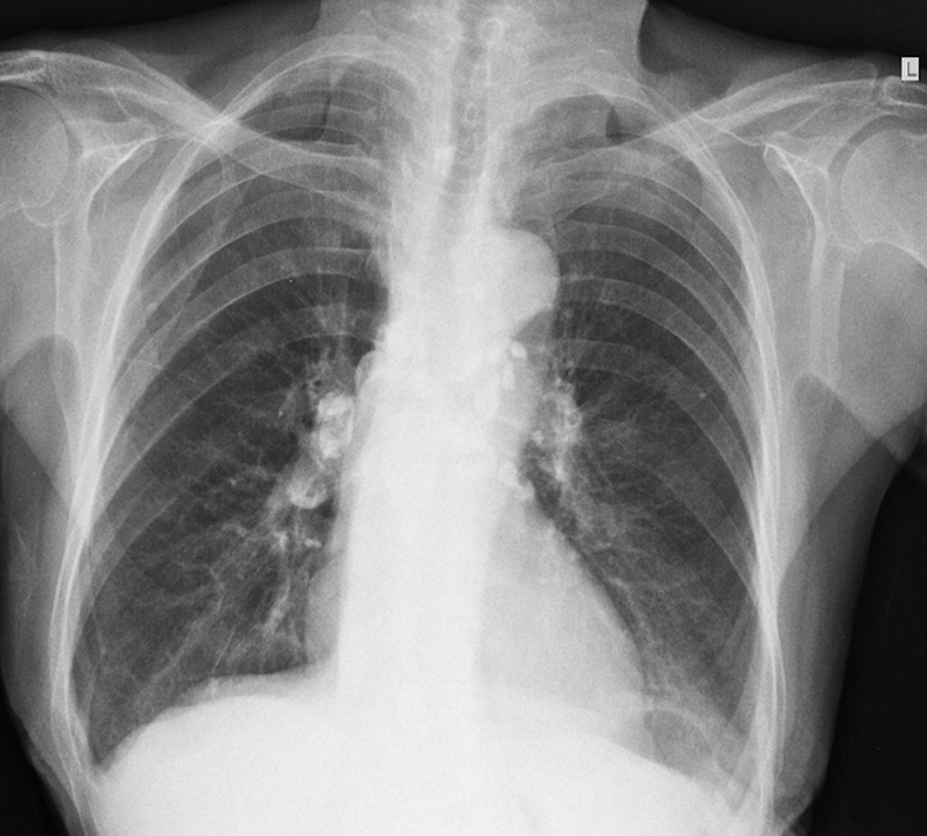
Primary Pulmonary Tuberculosis Ranke Complex Radiology Case Radiopaedia Org from prod-images-static.radiopaedia.org Pleural effusion on the same side is a common finding. Primary tuberculosis may affect any part in the lung. These unneeded regions would just make the extraction of the tuberculosis much more difficult and so we decided to simply pull out the lung regions. There is also suspicion of extrapulmonary tuberculosis, such as gastrointestinal or urogenital tuberculosis. The appearance is typical for chronic pulmonary tuberculosis but may also occur with chronic pulmonary histiocytosis and chronic pulmonary coccidioidomycosis. The cdc states that a positive test for tb infection only tells that a person has been infected with tb germs. It may be a primary tuberculous infection, secondary infection or appear as chronic scarring. Tb disease should be suspected in persons who have any of the following symptoms:
Chronic cough, fever, cough with bloody mucus, weight loss
Ct of the thorax is feasible if: The typical image is a homogeneous or heterogeneous white spot at the base of the affected lung or the middle third. The lungs are the most common site of primary infection by tuberculosis and are a major source of spread of the disease and of individual morbidity and mortality. The article reviews the imaging findings in ctb on various modalities. Thus, the differential diagnosis of pulmonary tuberculosis includes very many diseases. The main tb bacterium is mycobacterium tuberculosis (m. Diagram of a treatment algorithm for active tuberculosis. A positive tb skin test or tb blood test only tells that a person has been infected with tb bacteria. 37 yo with no pmhx with symptoms of cough, diarrhea, nausea and Abnormalities on chest radiographs may be suggestive of, but are never diagnostic of tb, but can be used to rule out pulmonary tb. Chest tuberculosis (ctb) is a widespread problem, especially in our country where it is one of the leading causes of mortality. And a discussion of other. Metrohealth and case western reserve university, affiliated since 1914, partners in advancing patient care through research and teaching.
Tb disease is treated by taking several drugs as recommended by a health care provider. There is also suspicion of extrapulmonary tuberculosis, such as gastrointestinal or urogenital tuberculosis. This helps your doctor choose the medications that are most likely to work. These unneeded regions would just make the extraction of the tuberculosis much more difficult and so we decided to simply pull out the lung regions. Less than 50% of adults with primary tuberculosis.
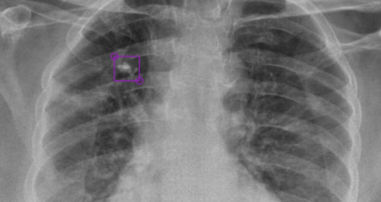
Deeptek Detects Tuberculosis From X Rays With Ai Nvidia Blog from blogs.nvidia.com Tb disease should be suspected in persons who have any of the following symptoms: This procedure is used as a secondary screening method in patients who have had a positive skin test and in patients who are at high risk for tuberculosis infection but have not had a positive skin test. 37 yo with no pmhx with symptoms of cough, diarrhea, nausea and In some cases, the patients may experience acute onset and high fever. Metrohealth and case western reserve university, affiliated since 1914, partners in advancing patient care through research and teaching. Abnormalities on chest radiographs may be suggestive of, but are never diagnostic of tb, but can be used to rule out pulmonary tb. An exudative lesion, a proliferative lesion, and a fibrotic lesion, and because it may invade all the structure. The lungs are the most common site of primary infection by tuberculosis and are a major source of spread of the disease and of individual morbidity and mortality.
Primary tuberculosis may affect any part in the lung.
Abnormalities on chest radiographs may be suggestive of, but are never diagnostic of tb, but can be used to rule out pulmonary tb. The samples are tested for tb bacteria. The article reviews the imaging findings in ctb on various modalities. The lungs are the most common site of primary infection by tuberculosis and are a major source of spread of the disease and of individual morbidity and mortality. Chest tuberculosis (ctb) is a widespread problem, especially in our country where it is one of the leading causes of mortality. This helps your doctor choose the medications that are most likely to work. Diagram of a treatment algorithm for active tuberculosis. We also attempt to categorize the findings into those definitive for active tb, indeterminate for disease activity, and those indicating healed tb. Tb disease should be suspected in persons who have any of the following symptoms: Less than 50% of adults with primary tuberculosis. An exudative lesion, a proliferative lesion, and a fibrotic lesion, and because it may invade all the structure. Chronic cough, fever, cough with bloody mucus, weight loss Pleural effusion on the same side is a common finding.


0 Komentar